What is a transesophageal echocardiogram?
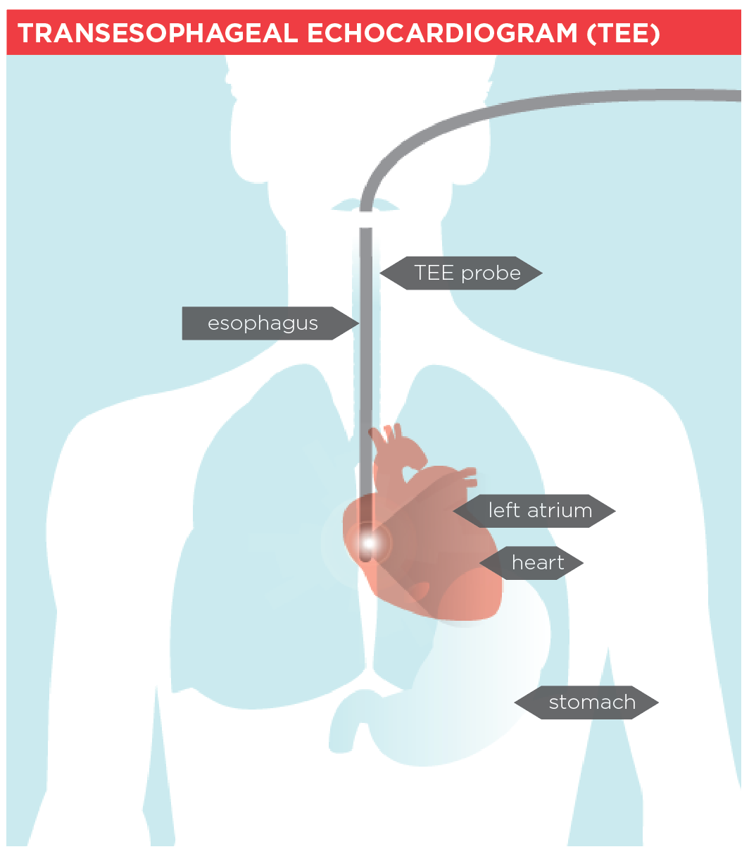
A transesophageal echocardiogram (TEE) is a special type of echocardiogram. Like an echocardiogram, the TEE uses high-frequency sound waves (ultrasound) to examine the structures of the heart. A transducer (a unit that directs the sound waves) is placed in the esophagus (the pipe that connects the mouth to the stomach). The esophagus is close to the heart, so images from a TEE can give very clear pictures of the heart and its structures.
Why it’s done
It is usually done when your doctor wants to look more closely at your heart to see if it could be producing blood clots.
Beat heart disease.
Join the fight to end heart disease and stroke.
How to prepare
- You will be asked not to eat or drink for 4 to 6 hours before your exam and to take any prescribed medications with only a sip of water.
- You should also arrange to have someone drive you home after the exam in case you are feeling drowsy.
What to expect
- You may be given a mild sedative to help you relax.
- You may also be given oxygen during the procedure.
- Your throat will be numbed with an anesthetic, then a flexible tube about the size of your index finger is inserted into your mouth and down your esophagus.
- During the procedure, you may feel the probe moving, but it won't be painful or interfere with your breathing.
- The transducer at the tip of the tube releases sound waves that bounce off your heart and are converted into pictures on a video screen.
- The doctor can move the tube up, down and sideways to look at different parts of your heart from different angles.
- The test usually takes about 20 to 40 minutes.
If you require more detailed information, check with the facility where you are having your exam.
